Mobile Tracking Device for Medical Applications
The Problem
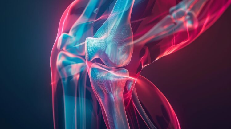
Prior to the use of fluoroscopy, kinematics of human joints were assessed using cadavers or motion analysis systems. Unfortunately, cadaver testing does not allow for in vivo soft-tissue conditions to be present, and motion analysis systems induce significant out-of-plane error due to skin motion. Fluoroscopy has become the gold standard for assessing in vivo human joint motion, but present-day fluoroscopic units are fixed to the ground. Therefore, those units are very limited, not allowing for continuous motion testing or testing of more complex daily activities.
The Solution
Researchers at the University of Tennessee have developed a first of its kind technology that integrates multiple sensing devices with a mobile robotic system so that specific skeletal joints of human subjects can be examined in real-time during natural ranges and rates of movement. An electrically driven mechanical arrangement would be provided to allow the entire mobile tracking device to unobtrusively track the human subject during normal movements.
A prototype implementation includes a separate tracking system that allows the x-ray source and imaging features of a fluoroscope to closely track the desired geometrical features of the selected skeletal joint. Through the use of this new technology, fluoroscopic image quality has been significantly advanced, because the unit now moves with the patient.
Therefore, patients can be evaluated performing their normal daily activities, and multiple activities can be assessed while the subject performs the motions at their normal speeds. The prototype has been used successfully in studying 300+ subjects.
Benefits
| Benefit |
|---|
| Applicable for knee, hip, and ankle skeletal joints. Extensions to shoulders and cervical spine are feasible. |
| Easy to view real-time movement and complete in vivo data acquisition. |
| First of its kind to move and track joints. |
| Opportunity for clinical diagnosis during weight-bearing, dynamic activities with the complete and natural engagement of a subject's musculoskeletal system. |
| System can also serve as a standard "static" fluoroscope for many clinical needs. |
More Information
- Kusum Rathore, Ph.D.
- UTRF Vice President
- 865-974-1882 | krathore@tennessee.edu
- UTRF Reference ID: 04016
- Patent Status: 8,406,845 B2
Innovators
William Hamel

Professor Emeritus, Department of Mechanical, Aerospace, and Biomedical Engineering (MABE), Tickle College of Engineering, UT Knoxville
Dr. Hamel received his PhD from the University of Tennessee. His research interests are in robotics and automation. He is a fellow of the Institute of Electrical and Electronic Engineers and the American Society of Mechanical Engineers.
Read more about William HamelRichard Komistek

Fred M. Roddy Professor, Department of Mechanical, Aerospace, and Biomedical Engineering Co-Director of Center for Musculoskeletal Research
Dr. Komistek received his PhD from the University of Memphis in 1992. His research interests include developing mathematical models of the human body musculoskeletal system, determination of in vivo mechanics of human joints, analysis of in vivo vibration and sound, and improving the quality of life for patients afflicted with arthritis.
Read more about Richard Komistek